|


|
|
Peyre, J-B., and Aigaki, T. 1999. A poor man's GFP desktop viewer.
Dros. Inf. Serv. 82: 97-98. View PDF
The Green Fluorescent Protein (GFP) is more and more widely used as a powerful tool for biologists, and especially drosophilists. One application that is unique to Drosophila is the construction of green balancers (Reichhart and Ferrandon, 1998). The advantages of using GFP expression as a reporter or as a dominant marker for balancer are clear; however, not everyone has an epifluorescence microscope in the laboratory, and even those who have one may feel inconvenienced to have to turn on the microscope every time one wants to observe a sample. Here is the description of the "GFP viewer", a Do-It-Yourself benchtop device designed for the observation of S65T modified GFP (Heim et al., 1995).
The GFP viewer can be used as a complement of an epifluorescence microscope or as a low-price/low-bulk alternative for the observation of S65T-GFP specimens. It allows observation and rapid sorting of GFP expressing bacterial clones, P{Gal4}/UAS-GFP or GFP balancer larvae, and can be used in combination with a standard binocular microscope. It can be turned on and off at will, when one needs to check for GFP expression of EGFP or S65T-GFP transgenic samples. It can be built easily, one needs just some soldering ability and common tools (screwdriver, drill); Its design is quite simple and illustrated by Figure 1:
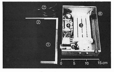 |
Figure 1. The GFP viewer. |
The elements needed for building the GFP viewer are:
– A set of polycarbonate blue and yellow color filters such
as those used by the lighting industry such as roscolux/supergel filters.
Two Glass or Plexiglas 200 ´ 145 mm rectangular plaques (2) are used to fix two
or three layers of blue filter on the box top. Putting the yellow filter (two
layers) into a stationery plastic holder (1) allows for handling it over the
observed samples. The blue filter is chosen to cut wavelength longer than 500 nm, whereas the yellow filter
eliminates wavelength shorter than 500 nm.
– A compact fluorescent lamp (3) with socket. It should emit around 480 nm. A Blue compact
fluorescent lamp should be preferred to the Daylight type.
– A ballast (4) for the neon tube and a starter lamp (5)
with socket; interrupter (6), fuse, cable and plug (7).
– A plastic, wood or metal box (8), preferably reflective
or white inside. Putting a rectangular mirror under the fluorescent lamp gives
a good increase in luminosity.
The two major elements, crucial for efficiency, the fluorescent lamp emission spectrum and the filter set transmission spectrums, have to be chosen carefully so as to fit the excitation/emission spectra of the S65T-GFP (Heim et al., 1995). When leaving the viewer on for long times, temperature buildup could occur, especially if using a daylight lamp. Ventilation has to be assured by drilling holes in the box or using metallic meshes, to allow air flow.
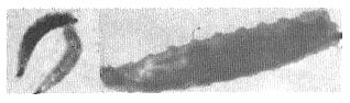 |
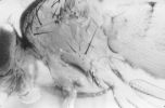
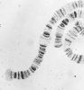
Figure 2. Observation of GFP expressing larvae: At left: 3rd instar larvae. upper: control, lower: Bloomington stock 4559 (FM7i-pAct-GFP). At right: P{Gal4} enhancer/UAS-GFP 3rd instar larva: GFP Expression is visible in the imaginal discs and salivary glands. |
Pictures (Figure 2) were taken using the GFP viewer and a Sony DKC5000 CCD (contrast and luminosity have been altered to accommodate grayscale images, originals visible online).
Using the GFP viewer, it is possible to observe the expression pattern and easily discriminate P{Gal4} enhancer trap/ UAS-GFP larvae due to the high expression of GFP. Sorting larvae that carry actin 5c-GFP balancers (Reichhart and Ferrandon, 1998) requires more attention as the pattern and level of expression may be lower in those larvae.
Inquiries for more technical details are also welcome by E-mail at peyre@c.metro-u.ac.jp or by mail at the laboratory. More figures and color pictures are available online at: http://www.sci.metro-u.ac.jp/~akira/gfpview.html. For people interested in buying an already-made version of the GFP viewer, a company will start producing it soon. We will be happy to send its name and address upon request.
Acknowledgment: J-B Peyre is supported by a scholarship from the Rotary Club - Yoneyama Foundation.
References: Heim, R., A.B. Cubitt, and R.Y. Tsien 1995, Improved green fluorescence. Nature 373: 663-664; Reichhart, J-M, and D. Ferrandon 1998, Greens Balancers. Dros. Inf. Serv. 81: 201-202.