|


|
|
Rudolph, T., B. Lu, T. Westphal, J. Szidonya, J. Eissenberg, and G. Reuter. 1999. New type of CyO and TM3 green balancers.
Dros. Inf. Serv. 82:99-100. View PDF
For differentiation between mutant homo- or heterozygotes during different stages of development, recessive visible mutations can be used (Lindsley and Zimm, 1992). More recently, transgenes expressing dominant markers were introduced into balancer chromosomes, allowing identification of balancer hetero- or homozygotes throughout development. Reichhart and Ferrandon (1998) used the 5C actin gene promotor driving GFP expression to construct “green balancers”. In order to avoid the strong maternal contribution of GFP seen in crosses using these balancers, we constructed a new set of CyO and TM3 green balancers. With the help of P{w[+m]hsp70:GAL4} and P{w[+m]UAS:GFP} elements which had been adjusted to heat shock independent expression after a series of remobilization crosses, balancers were established which allow easy differentiation between mutant homo- and heterozygotes during early embryogenesis, throughout all larval stages, in pupae and in adults. Finally, a series T(Y;2)CyOGFP, T(Y;3)TM3GFP and T(2;3)CyOGFP-TM3GFP translocations have been isolated. The T(2;3)CyOGFP-TM3GFP translocations combine the staining pattern of both CyOGFP and TM3GFP and are especially useful for genetic analysis.
The P{w[+m]UAS:GFP} element expresses the natural form of GFP (Yeh et al., 1995), allowing GFP detection under normal UV microscope with DAP1 filter. In our experiments, we use a Zeiss UV dissecting microscope. For analysis of embryos (without dechorionation) and early first instar larvae a regular fluorescence microscope can be used.
The green balancers were constructed by the following steps:
(1) Jump of P{w[+m]UAS:GFP} onto CyO and TM3, Ser: an X chromosomal P{w[+m]UAS:GFP} insert was remobilized in males of the constitution P{w[+m]UAS:GFP}/Y; CyO/+; P{(ry[+])2-3} Sb/+ and P{w[+m]UAS:GFP}/Y; P{(ry[+])2-3} Sb/TM3, Ser males, respectively. P{ry[+])2-3} is described in Robertson et al. (1988). Individual F1 w[+m] CyO or w[+m] TM3 males were tested for linkage of w[+m] with the dominant Cy and Ser marker mutations, respectively. From the set of about 10 to 15 CyO, P{w[+m]UAS:GFP} and TM3, P{w[+m]UAS:GFP} chromosomes, representative lines expressing a homogenous yellowish eye color were selected for further work.
(2) Jump of P{w[+m]hsp70:GAL4} onto CyO, P{w[+m]UAS:GFP} and TM3, P{w[+m]UAS:GFP}: a phenotypically nearly red eye second chromosomal P{w[+m]hsp70:GAL4} and third chromosomal P{w[+m]hsp70:GAL4} line was obtained from J. Urban and G. Technau (University of Mainz, Germany). Males of the genotypic constitution P{(ry[+])2-3}/Y; CyO P{w[+m]UAS:GFP}/ P{w[+m]hsp70:GAL4} and P{(ry[+])2-3}/Y; TM3, y[+] Ser P{w[+m]UAS:GFP}/ P{w[+m]hsp70:GAL4} were crossed to w/w females. In the F1 generation, Cy or Ser males expressing a wild type red eye color were collected. Linkage of the red eye color phenotype with Cy or Ser dominant markers were tested by a backcross to w/w females. Balancers were selected which gave strong green fluorescence after heat shock treatment. In order to select for heat shock-independent expression of green fluorescence, further remobilizations were performed.
(3) For remobilization of P{w[+m]hsp70:GAL4 on CyO, P{w[+m]hsp70:GAL4} P{w[+m]UAS:GFP} and TM3, Ser P{w[+m]hsp70:GAL4} P{w[+m]UAS:GFP} balancer-bearing females were crossed to TM3, ryRK Sb e P[(ry+)2-3]/Df(3R)C4 (Reuter et al., 1993) or Sb P[(ry+)2-3]/TM6 males, respectively. Offspring males w/Y; CyO, P{w[+m]hsp70:GAL4} P{w[+m}UAS:GFP}/+; TM3, ryRK Sb e P[(ry+)2-3]/+ and w/Y; TM3, Ser P{w[+m]hsp70:GAL4} P{w[+m]UAS:GFP}/Sb P[(ry+)2-3] were crossed to w/w females. Flies were allowed to lay eggs on small petri dishes, which were inspected for freshly hatched first instar larvae with strong GFP fluorescence. Individual larvae were collected and grown to adults. From those CyO, P{w[+m]hsp70:GAL4} P{w[+m]UAS:GFP}/Sco and TM3, Ser P{w[+m]hsp70:GAL4} P{w[+m]UAS:GFP}/Sb stocks were constructed. Finally, chromosomes were selected which expressed heat shock-independent green fluorescence beginning in early embryogenesis and continuously throughout the rest of development.
(4) We also isolated a series of CyGFP-TM3GFP, Y-CyOGFP, Y-TM3GFP and Y-CyOGFP-TM3GFP translocations after irradiation of w/Y; CyOGFP/+; TM3GFP/+ males with 4000R of X-rays. The irradiated males were crossed to w/w females and F1 w/Y; CyOGFP/+; TM3GFP/+ and male offspring tested for linkage between Cy and Ser or Cy and Ser and the Y chromosome, respectively, after backcrossing to w/w females. From 4717 males tested, 160 translocations were identified (142 T(2;3)CyOGFP-TM3GFP, 4 T(Y;2)CyGFP; 7 (T(Y;3)TM3GFP and 7 T(Y;2;3)CyGFP-TM3GFP). From these, representative translocations were selected, inspected for GFP expression and strains constructed.
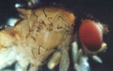
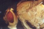
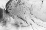
Expression Pattern of GFP: Strongest GFP expression is seen in T(2;3)CyOGFP-TM3GFP. None of the balancers show any maternal contribution of GFP, and mutant homo- and heterozygotes can be distinguished from the stock in ca. 10-12 hour old embryos (without dechorionation). The most pronounced fluorescence is visible in salivary glands and the midgut region. In addition to the strong fluorescence of salivary glands and midgut, a more homogenous background fluorescence is also found (e.g., imaginal discs, brain, fat body and gut) throughout larval stages of development. Heat shock treatment strongly enhances GFP expression, but even at 18oC the T(2;3)CyOGFP-TM3GFP translocation allows unambigous differentiation of balancer-bearing animals. We have successfully used these balancers in determining the lethal phase of recessive lethal PEV modifier mutations.
Selected pictures showing GFP expression in embryos and larvae can be viewed at http://www.biologie.uni-halle.de/Genetics/Drosophila/GreenBalancers/index.html.
The following stocks were sent to Umea and Bloomington stock centers:
CyOGFP: w[1]; CyO, P{w[+m]hsp70:GAL4} P{w[+m]UAS:GFP}/Sco[1]
TM3GFP: w[1]; TM3, y[+] ri[1] p[p] bx[34e] e[s] Ser[1] P{w[+m]hsp70:GAL4}
P{w[+m]UAS:GFP}/Sb[1]
T(2;3)CyOGFP-TM3GFP: w[1]; Sco[1]; T(2;3)CyO, P{w[+m]hsp70:GAL4} P{w[+m]UAS:GFP};
TM3, y[+] ri[1] p[p] bx[34e] e[s] Ser[1] P{w[+m]hsp70:GAL4} P{w[+m]UAS:GFP}/Sb[1].
Other translocation lines are available upon request.
References: Lindlsey, D.L., and G.G. Zimm 1992, In: The genome of Drosophila melanogaster. Academic Press, San Diego, CA.; Reichhart, J.M., and D. Ferrandon 1998, Dros. Inf. Serv. 81: 201-202; Reuter, G., G. Hoffmann, R. Dorn, and H. Saumwever 1993, Dros. Inf. Serv. 72: 78-79; Robertson, H.M., C.R. Preston, R.W. Phillis, D. Jonson-Schlitz, W.K. Benz, and W.R. Engels 1988, Genetics 118: 461-470; Yeh, E., K. Gustafson, and G.L. Boulianne 1995, Proc. Natl. Acad. Sci. USA 92: 7036-7040.