Ault, J.G. 2002. Improved ultrastructure of Drosophila tissue using high-pressure freezing and freeze substitution. Dros. Inf. Serv. 85: 126-127.
|
|
|
|||
|
|
||||
Improved ultrastructure of Drosophila tissue using high-pressure freezing and freeze substitution.
Ault, J.G. Division of Molecular Medicine, Wadsworth Center, New York State Department of Health, Albany, NY 12201-0509 USA. Correspondence: ault@wadsworth.org.
|
|
|
|
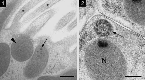
Figure 1. Mitochondria at the base of microvilli in a gut epithelial cell. The mitochondria are dense with matrix granules (arrow) and closed cristae without an open lumen (arrowhead). Actin bundles (asterisks) are seen inside the microvilli. Bar equals 0.2 µm. |
Figure 2. The axoneme and nucleus (N) in a spermatid. The axoneme displays the characteristic 9+2 cartwheel microtubular configuration with attached outer accessory microtubules (arrow). Bar equals 0.2 µm. |
Drosophila larval or adult tissue was packed with yeast paste into the specimen holders designed by Craig et al. (1987) and quickly frozen using a Balzers HPM 010 high-pressure freezer. The tissue was fixed by FS with 1% osmium tetroxide in acetone for 72 hours at -90oC, then 48 hours at -60oC. It was then washed several times at room temperature in 100% acetone and embedded in Epon/Araldite. Thin sections were cut and stained with uranyl acetate and lead citrate. Micrographs were taken using a Philips 301 or Zeiss 910 transmission electron microscope.
Signs of good preservation were straight microtubules, smooth membranes, full mitochondria, and abundant ribosomes both on the rough endoplasmic reticulum and in the full cytoplasm (Figures 1 and 2). The shape of the nucleus was similar to that observed in living cells using phase-contrast microscopy. Contrast can be increased by adding tannic acid or uranyl acetate to the FS solution. We believe that the improved preservation with HPF/FS allows the best chance to identify subtle changes in cellular structure caused by mutation in Drosophila. Though high-pressure freezers are expensive (around $185,000), a number of nationally funded EM centers have them, which can be used by outside scientists.
Acknowledgments: This work was done at the Wadsworth Center’s EM Core facility.
References: Craig, S., J.C. Gilkey, and L.A. Staehelin 1987, J. Microsc. 48: 103-106; McDonald, K., and M. K. Morphew 1993, Microsc. Res. Tech. 24: 465-473.