Dimitri, Patrizio, Fabrizio Rossi, and Nicoletta Corradini. 2002. FISH with rhodamin-labeled probes to Drosophila melanogaster chromosomes. Dros. Inf. Serv. 85: 117-118.
|
|
|
|||
|
|
||||
FISH with rhodamin-labeled probes to Drosophila melanogaster chromosomes.
Dimitri, Patrizio, Fabrizio Rossi, and Nicoletta Corradini. Dipartimento di Genetica e Biologia Molecolare "Charles Darwin", Università di Roma "La Sapienza", Piazzale A. Moro 5, 00185 Roma, Italy; Patrizio.Dimitri@uniroma1.it
Biotin-labeled
or digoxigenin-labeled probes coupled with secondary detection are routinely
used for fluorescence in situ hybridization
(FISH). However, FISH probes can be also labeled directly with fluorophores,
usually by incorporation of fluorescently conjugated nucleotides. Fluorescein-labeled
dNTP (green emission) or Cy3-labeled dUTP (red emission) are available from
several suppliers. We have prepared DNA probes labeled with tetramethylrhodamin-6
dUTP (red emission) using a rhodamin-nick translation kit (Boheringer, Mannheim).
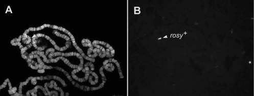
FISH with tetramethylrhodamin-6 dUTP labeled probes can be performed on both
polytene (Figure 1) and mitotic chromosomes of Drosophila melanogaster. The use of DNA probes directly labeled with this or with other
fluorescently conjugated nucleotides avoids the blocking and detection steps
and is particularly useful because it reduces the background and shortens
the procedure. It is important
to point out that the hybridization signal intensity obtained with tetramethylrhodamin-6
dUTP labeled probes on polytene chromosomes is comparable to that observed
using biotin-labeled or digoxigenin-labeled probes coupled with secondary
detection. On mitotic chromosomes, tetramethylrhodamin-6 dUTP labeled probes
may be not as efficient as biotin-labeled or digoxigenin-labeled probes in
revealing single-copy DNA signals, but it works efficently with BAC (bacterial
artificial chromosomes) DNA probes.
1. Label 1 mg of DNA probe (plasmids, BACs or PCR fragments) by Nick-translation using a rhodamin-nick translation kit (Boehringer, Mannheim).
2. Transfer the desired amount of labeled DNA into an eppendorf tube, add sonicated salmon sperm DNA (3 mg per slide) and dry in a savant centrifuge.
After the post-hybridization washes, slides incubated with probes directly labeled with tetramethylrhodamin-6dUTP or other fluorophores must be treated as follows:
1. Wash slides once for 5 min in 2´SSC 0.1% Tween 20 at room temperature.
2. Stain slides with 0.16 mg/ml 4,6-diamino-2-phenylindole-dihydrocloride (DAPI) dissolved in 2´SSC for 5 min at room temperature.
3. Rinse slides once in 2´SSC at room temperature, remove slides from 2´SSC and air dry.
4. Mount slides in 20mM Tris-HCl, pH 8, 90% glycerol containing 2.3% of DABCO antifade (1,4-diazo-bicyclo-(2,2,2)octane; Merck). Commercial antifade such as Vectashield H-1000 (Vector laboratories) can be also used. Seal coverslips with rubber cement and store at 4°C.
|
Figure 1. FISH localization of rosy+ gene on polytene chromosomes from the laboratory strain Charolles. A) DAPI staining; B) Fluorescent signal of the rosy + gene at 87D. The CAR 20 construct carrying the rosy marker was used as DNA probe. |