
Ault, J.G., and E. Shimakawa. 2002. A new virus-like particle found in Drosophila melanogaster. Dros. Inf. Serv. 85: 29-30.
|
|
|
|||
|
|
||||
A new virus-like particle found in Drosophila melanogaster.
Ault, J.G.,1 and E. Shimakawa2. 1Division of Molecular Medicine, Wadsworth Center, New York State Department of Health, Albany, NY 12201-0509 USA, ault@wadsworth.org; 2Biology Department, Chaminade University, Honolulu, HI 96816 USA, eshimaka@chaminade.edu
Reports of virus-like particles (VLPs) in Drosophila tissue, during the mid-1960s and early 1970s, were among the first evidence that viruses infect Drosophila melanogaster (Akai et al., 1967; Filshie et al., 1967; Philpott et al., 1969; Gartner, 1971; Felluga et al., 1971). Since then, only a few Drosophila viruses have been further characterized (Brun and Plus, 1980). Drosophila VLPs mostly appear oval to round in shape and form quasi-crystalline arrays, usually in the nucleus, but also in the cytoplasm. They range in average length from 400 to 727 angstroms and have been distinguished from one another according to size (Felluga et al., 1971).
 |
|
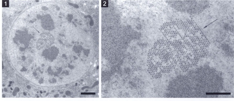
| Figure 1. A quasi-crystalline inclu-sion (arrow) of virus-like particles in the nucleus of a gut epithelial cell. Bar equals 1.0 µm. | Figure 2. Higher magnification of the inclusion in Figure 1. Thin fibers (arrow) are observed running along one side of the inclusion. The virus-like particles are hexagonally packed with spaces disrupting the periodicity. Bar equals 0.5 µm |
Acknowledgments: This work was done at the Wadsworth Center’s EM Core facility.
References: Akai, H., E. Gateff, L.E. Davis, and H.A. Schneiderman 1967, Science 157: 810-813; Brun, G., and N. Plus 1980, The Genetics and Biology of Drosophila (Ashburner, M., and E. Novitski, eds.) 2d, pages 625-702, Academic Press, New York; Felluga, B., V. Jonsson, and M.R. Liljeros 1971, J. Invert. Path. 17: 339-346; Filshie, B.K., T.D.C. Grace, D.F. Poulson, and J. Rehacek 1967, J. Invert. Path. 9: 271-273; Gartner, L.P., 1971, Experientia 27: 562-564; Philpott, D.E., J. Weibel, H. Atlan, and J. Miquel 1969, J. Invert. Path. 14: 31-38.