 | Spring Student Micrograph Contest - 2017-18 |  |
| Spring Student Micrograph Contest - 2017-18 | |
 |
 Petrographic Microscope Image: Quartz grains in clay, carbonate, and calcium sulfate matrix, Permian Redbeds, RB Core (2,743 m depth), Western Kansas. TEM Image: Jarosite alteration in jarosite + 20 wt.% CaCl2 flowing brine experiment -- high angle annular dark field STEM image |
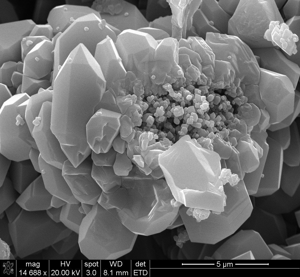
 SEM Image: Botryoidal mystery mineral (ongoing analyses) in Fe-bearing clay matrix, Permian Redbeds, RB Core (2,743 m depth), Western Kansas. |
 Prairie vole large intestinal section showing a goblet cell releasing mucin into the lumen containing bacteria. Tissue was fixed in a primary fixative of 2.5% glutaraldehyde in 0.1 M cacodylate buffer (pH 7.4) for 30-45 minutes. The samples were then secondarily-fixed in 2% osmium tetroxide with 0.1M cacodylate buffer (pH 7.4). The samples were washed with 0.05 M cacodylate buffer and en bloc stained with 20% ethanol (EtOH)/uranyl acetate (UA). The tissues were embedded in PolyBed resin. Gold-silver sections were double stained with lead citrate and uranyl acetate and examined with a Hitachi H-7000 electron microscope operated at 75kV. |
 Appendix for Micheal Anderson Description of Colon Nerve Mesh |
 Petalloid Anther: Arabidopsis thaliana plants with mutations in three NF-YC genes and a mutation in HY5 have anthers with petal characteristics (false colored yellow and white), a novel phenotype of these well studied mutants. This photograph was taken on the Zeiss Neon 40 Scanning Electron Microscope at 2.00 kV at a magnification of 150x. This image was false colored using GIMP image editing software. | |
 Euhedral microcrystalline quartz and micro-porous chert with examples for the micro- and nano- scale porosity present in the Carboniferous tripolitic chert reservoirs of the Midcontinent of the USA. The sample is from a roadcut in Pineville, Missouri |
 Plant penetration structure (appressoria) in anaerobic gut fungi, that looks like a pilot wheel. |
Please address any comments or inquiries about these Web pages to Scott Russell, snailmail: Samuel Roberts Noble Microscopy Lab, University of Oklahoma, Norman, OK 73019 (405) 325-4391 email: srussell@ou.edu