Pérez-Chiesa, I., and G. Rivera Colón. 2002. Morphology of mouth hooks during larval development of Drosophila dunni from Puerto Rico. Dros. Inf. Serv. 85: 28-29
PDF file
|
|
|
|||
|
PDF file
|
|
|||
Pérez-Chiesa, I., and G. Rivera Colón. 2002. Morphology of mouth hooks during larval development of Drosophila dunni from Puerto Rico. Dros. Inf. Serv. 85: 28-29.
Morphology of mouth hooks during larval development of Drosophila dunni from Puerto Rico.
Pérez-Chiesa, I., and G. Rivera Colón. Department of Biology, University of Puerto Rico, Río Piedras, P.R. 00931
The mouth hooks of Drosophila larvae are transitory structures, which may change their morphology with every molt. There is also much interspecies variation in morphology. The hooks may have zero, few or numerous teeth, which in turn, vary in size (Alpatov, 1929; Okada, 1963). Because the number of teeth present in the hooks from one instar to the other does not overlap (Alpatov, 1929), the morphology of the mouth hooks has been used to stage larval development in Drosophila melanogaster and other species (Bodenstein, 1950; Quintana and Juan, 1993; Amador and Juan, 1998). Behavioral and developmental studies with D. dunni from Puerto Rico, a member of the cardini group, require the accurate staging of larvae. Thus, we have examined the morphology of the mouth hooks during larval development.
|
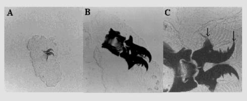
Figure 1. Larvae were either dissected or
squashed between a slide and a coverslip in a drop of glycerol and
viewed under a Nikon microscope at 400´. A, First instar larvae;
B, Second instar
larvae; C, Third instar larvae. |
Flies were allowed to lay eggs for periods of 30 minutes, on banana agar-medium seeded with live yeast to induce egg laying. Cultures were kept in an incubator at 25˚C and synchronized by removing all larvae that were present in the medium 24 hours later. The mouth hooks of first and second instar larvae were either dissected or obtained from the media during molting. Mouth hooks from third instar larvae in their wandering stage were not dissected but cleared by boiling in KOH for 3 minutes. All samples were mounted in glycerol and the hooks were measured from the tip to the base with an ocular micrometer as indicated by the arrows in Figure 1C. Most eggs hatched at 26.5 ± 0.5 hrs. The first, second and third instars last approximately 22 hrs, 28 hrs, and 74 hrs, respectively. The first molt occurs at 49 ± 1 hr after egg laying (AEL); the second molt at 77 ± 2 hr AEL. Pupation starts at 151 hr AEL. There can be much variation at this stage unless larvae are synchronized during the second molt. The adult emerges 12 days after egg laying. The mouth hooks of first instar larvae are small (26 ± 1.47 µm) with one or two sharp teeth in the middle of the hook (Figure 1A). Most larvae have only one tooth. Mouth hooks of second instar larvae are nearly double in size (44 ± 1.83 µm) and have only one prominent sharp tooth preceded by two to four middle sized teeth (Figure 1B). Third instar larvae have 6 to 12 teeth showing a more complex pattern. The size of the teeth increases with age. At the wandering stage, most larvae have two prominent sharp teeth with several middle-sized teeth between them, and in front of the first sharp tooth, towards the tip of the hook, there are several minute teeth (Figure 1C). The size of the hooks is 81 ± 7.45 µm. As in D. melanogaster and other species, the number of teeth in the mouth hooks of D. dunni showed no overlap from one instar to the next.
Acknowledgments: The work was supported by RISE-NIGMS
References: Alpatov, W.W.,1929, J. Exp.
Zool. 52: 407-437; Amador, A.,
and E. Juan 1998, Dros. Inf. Serv. 81: 153-154; Bodenstein, D., 1950,
In: Biology of Drosophila.
(Demerec M., ed.). John Wiley and Sons, p. 275; Okada, T., 1963, Evolution
17: 84-98; Quintana, A., and
E. Juan 1993, Dros. Inf. Serv. 72: 88.